Securing the airway in newborns with cleft lip or palate demands meticulous planning, precise technique, and clear visualization. Traditional adjuncts protect soft tissues only indirectly, leaving the palatal and alveolar regions susceptible to contact and shear. A customized protective palatal obturator aims to directly shield these structures during laryngoscopy and endotracheal tube passage while maintaining line-of-sight and access. The concept is simple but potentially consequential: place a contoured, low-profile barrier where contact is most likely and make it easy to insert and remove without interrupting the sequence of care.
Recent randomized evidence explores this approach during primary cheiloplasty, focusing on feasibility, safety, and procedural performance. The device is designed to integrate into existing workflows without creating new bottlenecks or compromising first-pass success. What follows reviews the clinical rationale, device design and deployment, and how such an adjunct might fit into broader pathways for neonatal cleft surgery and perioperative airway protection.
Protecting the neonatal airway in cleft surgery
Among high-stakes pediatric scenarios, Neonatal Intubation during cleft lip repair is both common and uniquely nuanced. Infants with Cleft Lip And Palate present with discontinuities of soft and hard tissues that alter the geometry of mask seal, blade placement, and tube advancement. In this setting, even gentle contact can translate to focal abrasion or pressure on fragile palatal edges or alveolar ridges. A protective obturator aims to offload that contact onto a smooth, custom-fitted interface while preserving the choreography of Airway Management. The device conceptually extends existing paradigms by adding anatomic specificity rather than replacing standard laryngoscopy or tube selection.
Clinical teams already balance visualization, speed, and tissue protection, especially when preoxygenation windows are short. The obturator approach seeks to augment that balance by lining potential contact points with a preformed shield that does not obstruct the view. By using the infant's own anatomy as the template, a custom device can seat predictably and resist displacement during Laryngoscopy. Importantly, the tool is intended to be neutral with respect to operator skill level, complementing familiar techniques across providers. If proven safe and practical, such an adjunct could help standardize steps that are otherwise heavily operator-dependent.
Why cleft anatomy complicates intubation
In infants with unilateral or bilateral clefts, absent or displaced palatal support leaves margins vulnerable to compressive and shear forces during blade insertion and tube delivery. Discontinuities can also create irregular channels that divert the endotracheal tube, subtly changing the angle needed for smooth passage. Mask ventilation may be less effective due to air leak at the cleft site, shortening the time available for secure intubation. These realities elevate the premium on efficient visualization and minimize tolerance for aborted attempts. A purpose-built barrier that shields the defect margins can reduce incidental contact while allowing standard physiology-driven anesthetic plans to proceed.
Beyond mechanical concerns, bleeding from minor abrasion can quickly obscure landmarks in a small field, compounding difficulty. A protective layer that smooths the palatal contour may reduce opportunities for contact-induced oozing and keep the laryngoscopic view cleaner. The device must accomplish this without adding bulk that narrows the working space or requires atypical maneuvers. That constraint pushes design decisions toward thin, contoured forms that maintain a stable seat. In effect, the goal is to provide a protective footprint that is physiologically and procedurally quiet.
Defining outcomes that matter to teams and families
For any adjunct in neonatal airway care, priority outcomes include protection of mucosa, maintenance of visualization, and preservation of first-pass success. Procedural efficiency and absence of device-related adverse events are also essential. Families value painless recoveries, and while most tissue contacts are minor, cumulative trauma can affect feeding comfort or early postoperative care. From a systems perspective, scalable solutions should work across varied provider experience, equipment brands, and operating room environments. Capturing these outcomes consistently supports reproducibility and real-world utility rather than single-center optimization.
The current RCT framework emphasizes clinically salient measures such as frequency of contact with palatal tissues, visible mucosal changes, ease-of-view grading, and need for additional maneuvers. It also treats workflow endpoints like setup time and removability as integral safety signals. This aligns with a growing emphasis on Patient Safety in pediatric surgical pathways, where low-frequency but high-impact events drive quality metrics. If protective obturators can reduce minor but consequential interruptions, they could compound benefits across the perioperative course. Such framing is necessary to evaluate a device whose value lies in preventing problems more than in solving them once present.
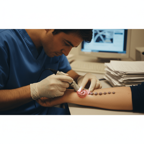
From impression to insertion: how the obturator works
Conceptually, the obturator is a custom-fitted palatal shield fabricated from a smooth, biocompatible material tailored to neonatal dimensions. The workflow begins with a gentle impression that captures the cleft geometry without distorting soft tissues. The resulting model guides fabrication of a thin device that cups the palatal vault and spans the cleft margins, creating a protective surface for procedural contact points. During intubation, the obturator is seated before laryngoscopy and removed after the tube is secured, typically without altering the sequence or equipment. This is a classic interface solution grounded in Prosthodontics, ported into the operating room to protect at-risk tissues.
Such adaptation reflects a broader bridge between Craniofacial Surgery and anesthetic technique. Protective devices are most useful when they fade into the background of the procedure and impose minimal cognitive load. The best designs are intuitive in orientation, have reliable tactile feedback at seating, and resist dislodgement with routine manipulations. At the same time, they should be instantly removable if visualization is impeded or any resistance is felt during tube passage. This balance of stability and reversibility is the hallmark of an effective intraoperative adjunct.
Customization, materials, and fit
Custom devices must be thin and smooth to avoid occupying valuable intraoral working space. Biocompatible thermoplastics or medical-grade elastomers can achieve the needed combination of flexibility and resilience. Precision matters: a seat that conforms closely to palatal contours spreads contact forces over a larger area and reduces focal pressure. The cleft span should be bridged by the device without transmitting force to the edges. In practice, small contour changes can make a large difference in how the obturator behaves under load.
Impression-taking in neonates requires gentle technique, minimal material volume, and rapid set times. Teams accustomed to alveolar molding or feeding obturators already manage these considerations safely. The protective obturator differs in its procedural role and duty cycle, facing brief but potentially more concentrated forces during laryngoscopy. Developers should specify temperature tolerance for sterilization pathways and recommend inspection criteria for surface integrity. A simple check-list aids repeatable quality before every use.
Workflow integration in the operating room
Integration starts with team briefing: when to seat the device, who confirms seating, and what to do if the view is suboptimal. The obturator is placed after induction but before blade entry, with a quick visual confirmation of position. The laryngoscope then proceeds as usual, with careful observation for displacement or unexpected resistance. If visualization is compromised, the plan should include immediate removal and a return to baseline technique. Clear roles and short verbal checks keep the sequence predictable.
Device removal follows tube confirmation and securing, minimizing the chance of dislodgement by tube motion. Simple retention features, like subtle flanges that sit outside the cleft margin, can provide tactile confirmation during insertion and easy grasp for removal. It is important that laryngoscope blade choice, tube size, and stylet use remain at the discretion of the anesthesiologist. The adjunct should not force a change in familiar tools or techniques. Minimalism in design and workflow is the best path to adoption.
Safety checks and troubleshooting
Pre-use inspection focuses on surface smoothness, absence of burrs, and confirmation of correct patient identity and orientation. During insertion, the device should seat without resistance and remain stable under gentle tongue depression. If any rocking is observed, reseating or switching to baseline technique is prudent. Under no circumstance should the device be forced into place or used to lever tissues. The default is always to prioritize visualization and first-pass success over persistence with the adjunct.
Signs that warrant removal include partial obstruction of view, increased force requirement, or audible clicking that suggests contact at an edge. The anesthesiologist should feel comfortable abandoning the device if conditions are not improved or are even slightly worse. Post-use checks can document whether contact marks are present on the obturator surface rather than on mucosa, supporting the mechanism of action. Documentation of any deviation builds a safety profile over time. A conservative troubleshooting algorithm protects both patient and operator.
Evidence, adoption, and next steps
The emerging evidence base includes a Randomized Controlled Trial in infants undergoing primary cleft lip repair, assessing feasibility, safety, and goal-concordant procedural metrics. The RCT, indexed at PubMed, provides a structured look at whether a protective palatal obturator can integrate smoothly into existing practice. Outcomes of interest include visible soft tissue changes, ease-of-view scores, need for ancillary maneuvers, and time to intubation. While quantitative details are best read directly in the report, the design highlights practical endpoints that translate readily into operating room decisions. This is a useful step beyond descriptive case experience toward standardized evaluation.
For adoption, context matters. Centers with established cleft pathways and in-house dental or prosthetic collaboration will likely find fabrication and maintenance straightforward. Training curves are modest if the design is intuitive and the seating step is brief. Institutions without ready access to fabrication can consider regional partnerships or small-batch kits sized by common anthropometric ranges. Ultimately, implementation depends on convincing evidence that the adjunct reduces contact-related sequelae without compromising speed or view.
Trial design and what was measured
In perioperative innovation, defining what to measure is often as important as the intervention. The trial captured procedural events that map to clinician priorities, including mucosal contact markers, laryngoscopic grade of view, tube passage ease, and any adjunct-related interruptions. Safety signals comprised device dislodgement, obstruction, or need for abandonment. Workflow measures like placement time and removability were recorded to ensure net neutrality or improvement. These choices align with quality and safety initiatives in pediatric surgery and anesthesia.
Equally important is how data are collected. Prospective checklists, standardized grading scales, and immediate post-procedure inspections limit recall bias and apply a common language to subtle events. Early studies of device adjuncts can fall prey to enthusiasm bias; rigorous, prespecified measures help mitigate that risk. In reporting, clarity about per-protocol versus intention-to-treat analysis supports reproducibility. Although effect sizes will evolve with broader adoption, the initial framework sets a durable template for subsequent studies.
Who benefits most and when to avoid use
Patients with broader palatal spans or friable mucosa may stand to benefit most from barrier protection. Conversely, if very limited oral aperture or atypical anatomy impedes seating, defaulting to baseline technique is sensible. The adjunct is not a substitute for operator judgment or for alternate airway strategies if visualization is poor. Teams should maintain readiness for mask ventilation, supraglottic placement, or fiberoptic assistance according to standard algorithms. A clear stop rule prevents prolonging attempts in conditional benefit scenarios.
Operator factors also matter. Providers early in training can use the obturator as a protective backdrop, but supervision should ensure that view quality remains uncompromised. Experienced clinicians may appreciate the adjunct most in borderline cases where minimal contact would otherwise be unavoidable. In all cases, the device is best framed as an optional layer of safety rather than a requirement. Transparent criteria for when not to use the device make the pathway more robust.
Implementation roadmap and research agenda
Implementing a protective obturator pathway can proceed in phased steps. Start with simulation using neonatal mannequins and cleft models to refine seating and removal choreography. Move to a pilot with carefully selected cases, collecting the same endpoints used in clinical trials. Include structured debriefs to capture ergonomic observations and refine device design or instructions for use. A brief competency checklist helps anchor training for rotating staff. Early successes and candor about limitations build trust and momentum.
The research agenda should prioritize multicenter validation, longer-term outcomes, and standardized definitions of contact-related events. Subgroup analyses by cleft phenotype and operator experience can uncover where benefit is greatest. A registry model could accelerate learning and guide iterative design improvements. Collaboration between anesthesiology, surgery, and dental teams ensures that device refinements track real-world needs. Over time, harmonizing metrics will make cross-study comparisons meaningful and accelerate consensus.
Integrating the obturator into broader safety bundles may amplify impact. Such bundles could include pre-induction briefings, equipment readiness checks, and standardized grading of view and contact. Reporting near misses and small injuries transparently keeps attention on continuous improvement. Institutions should also consider procurement pathways that ensure consistent material quality and sterilization compatibility. The aim is to make the right thing the easy thing for every team, every time.
Looking ahead, protective interfaces that respect neonatal anatomy and preserve procedural flow represent a promising direction for airway care. The customized palatal obturator is emblematic of this approach: it is small, targeted, and designed to get out of the way. Evidence from randomized evaluation is a strong start, but broader validation and device iteration will determine staying power. The opportunity is to reduce the invisible toll of minor contacts while keeping first-pass success and visualization sacrosanct. That balance is the heart of innovation in Pediatric Anesthesia and Device Innovation.
LSF-2697470695 | October 2025
How to cite this article
Team E. Customized palatal obturator for safer neonatal intubation. The Life Science Feed. Published November 5, 2025. Updated November 5, 2025. Accessed April 2, 2026. https://thelifesciencefeed.com/genetics/genetic-diseases-inborn/innovation/customized-palatal-obturator-for-safer-neonatal-intubation.
Copyright and license
© 2026 The Life Science Feed. All rights reserved. Unless otherwise indicated, all content is the property of The Life Science Feed and may not be reproduced, distributed, or transmitted in any form or by any means without prior written permission.
Fact-Checking & AI Transparency
This content was produced with the assistance of AI technology and has been rigorously reviewed and verified by our human editorial team to ensure accuracy and clinical relevance.
References
- Customized protective palatal obturator for intubation in newborns in cleft lip surgery: a randomized controlled trial. https://pubmed.ncbi.nlm.nih.gov/40981509/.
Related Articles