Urine microscopy, a cornerstone of laboratory medicine for decades, faces a concerning decline in utilization. This isn't merely a matter of technological obsolescence; it reflects deeper systemic issues within healthcare. We're seeing a confluence of factors, from shifting reimbursement models to evolving training priorities, that collectively devalue this essential diagnostic skill. The downstream effects on patient care and healthcare expenditure warrant a closer look.
Hospital administrators and program directors need to recognize that losing this skill has consequences. When was the last time your institution audited the competency of urine microscopy among its lab staff and residents? Are you truly prepared to handle complex renal cases with increasingly limited diagnostic tools?
Clinical Key Takeaways
lightbulb
- The PivotThe historical reliance on urine microscopy is threatened by financial disincentives and reduced training opportunities, jeopardizing diagnostic capabilities.
- The DataStudies have shown a correlation between decreased microscopy utilization and potential increases in downstream testing and healthcare costs due to missed or delayed diagnoses.
- The ActionHospital administrators should re-evaluate laboratory reimbursement models and advocate for enhanced medical education to maintain competency in urine microscopy.
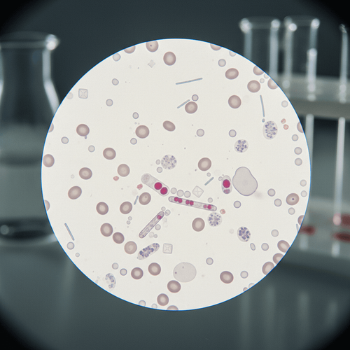
The slide on which urine microscopy rests is gathering dust in many modern labs. While automated urine analyzers offer speed and convenience, the human eye, when properly trained, often detects subtleties that machines miss. The question isn't whether automation has a role- it certainly does- but whether we are sacrificing diagnostic accuracy and long-term cost-effectiveness by prematurely phasing out a valuable skill.
The Economic Squeeze on Microscopy
One of the primary drivers behind the decline is, unsurprisingly, financial. Fee-for-service reimbursement models often favor high-volume, automated tests over labor-intensive manual procedures like microscopy. Laboratories face constant pressure to cut costs and increase throughput. A urine dipstick read by an automated analyzer generates revenue quickly and efficiently. A trained pathologist or lab technician spending valuable time meticulously examining a urine sediment? That's seen as a drag on the bottom line.
This economic pressure has a direct impact on staffing. Hospitals and private labs alike are less willing to invest in specialized training for urine microscopy, leading to a gradual erosion of expertise. The Centers for Medicare & Medicaid Services (CMS) reimbursement rates for manual microscopy have not kept pace with the cost of labor and supplies, further incentivizing the shift towards automation. This is a classic example of short-term cost savings leading to potentially higher long-term expenses, as missed or delayed diagnoses necessitate more complex and costly interventions later on.
Regulatory Burden and CLIA
The Clinical Laboratory Improvement Amendments (CLIA) regulations, while intended to ensure quality and accuracy, can inadvertently create barriers to manual microscopy. Meeting CLIA requirements for proficiency testing and quality control adds to the administrative burden and cost of maintaining a fully functional microscopy service. Smaller labs, in particular, may find it challenging to comply with these regulations, leading them to abandon manual microscopy altogether. There's a perception- whether justified or not- that automated analyzers are easier to validate and maintain under CLIA guidelines.
The Training Gap in Microscopy
Perhaps the most insidious threat to urine microscopy is the decline in formal training opportunities. Many residency programs, particularly in internal medicine and family medicine, now place less emphasis on hands-on microscopy experience. The curriculum is already packed with competing demands, and urine microscopy is often seen as a lower-priority skill compared to, say, interpreting complex imaging studies or managing chronic diseases. This creates a vicious cycle: as fewer clinicians and lab technicians are trained in microscopy, the expertise needed to teach the next generation dwindles, perpetuating the decline.
This contradicts the 2021 Kidney Disease Improving Global Outcomes (KDIGO) guidelines which continue to recommend microscopic examination as a crucial component of evaluating kidney diseases. The KDIGO guidelines emphasize the importance of identifying specific elements in urine sediment to guide diagnosis and treatment, a task that automated analyzers often cannot perform with sufficient accuracy.
Is it Really More Accurate?
That's the core question, isn't it? Proponents of automation argue that modern urine analyzers are highly sensitive and specific, capable of detecting most clinically significant abnormalities. However, these analyzers often rely on algorithms and pre-set thresholds that may not be appropriate for all patient populations. Furthermore, they can miss subtle findings, such as dysmorphic red blood cells or certain types of casts, that are crucial for diagnosing specific renal diseases. A systematic review comparing manual microscopy to automated analysis found that while automation had high sensitivity for detecting leukocytes and nitrites, its specificity for detecting casts and crystals was significantly lower. False negatives can lead to delayed diagnoses and inappropriate treatment, while false positives can trigger unnecessary and costly investigations.
The Hidden Costs of Automation
The allure of automation lies in its apparent efficiency and cost-effectiveness. However, a closer examination reveals several hidden costs. First, automated analyzers often generate a high number of “equivocal” or “uninterpretable” results, requiring manual review and reflex testing. This can negate the purported time savings of automation and increase overall laboratory workload. Second, the reliance on automated results can lead to over-testing and inappropriate use of antibiotics. For example, a positive leukocyte esterase result on an automated analyzer may prompt a clinician to prescribe antibiotics for a urinary tract infection, even in the absence of significant symptoms or microscopic evidence of infection. This contributes to the growing problem of antibiotic resistance and increases healthcare costs.
Finally, consider the cost of maintaining and upgrading automated analyzers. These machines require regular maintenance, calibration, and software updates, all of which add to the overall cost of ownership. Smaller labs may find it difficult to afford these expenses, forcing them to rely on older, less accurate equipment.
The decline in urine microscopy carries significant financial and workflow implications for health systems. The increased reliance on automated urinalysis often leads to more frequent and costly reflex testing to confirm or refute initial findings. This additional testing not only increases laboratory expenses but also adds to the workload of already stretched lab personnel. Furthermore, the potential for missed or delayed diagnoses due to the limitations of automated analysis can result in higher downstream healthcare costs associated with managing complications and prolonged hospital stays. Hospital administrators should re-evaluate their laboratory budgets and consider investing in training programs and updated equipment to maintain competency in urine microscopy. Advocating for more appropriate reimbursement models that recognize the value of manual microscopy is also essential. Failure to address these systemic issues will only exacerbate the problem and ultimately compromise patient care.
LSF-2990891313 | December 2025

How to cite this article
O'Malley L. The systemic undermining of urine microscopy. The Life Science Feed. Published December 31, 2025. Updated December 31, 2025. Accessed April 1, 2026. https://thelifesciencefeed.com/nephrology/acute-kidney-injury/policy/the-systemic-undermining-of-urine-microscopy.
Copyright and license
© 2026 The Life Science Feed. All rights reserved. Unless otherwise indicated, all content is the property of The Life Science Feed and may not be reproduced, distributed, or transmitted in any form or by any means without prior written permission.
Fact-Checking & AI Transparency
This content was produced with the assistance of AI technology and has been rigorously reviewed and verified by our human editorial team to ensure accuracy and clinical relevance.
References
- Fogazzi, G. B., Ponticelli, C., & Ritz, E. (2001). The Kidney in Systemic Diseases. In: Massry SG, Glassock RJ, eds. Textbook of Nephrology (4th ed.). Lippincott Williams & Wilkins.
- KDIGO. (2021). KDIGO 2021 Clinical Practice Guideline for the Management of Glomerular Diseases. Kidney International Supplements, 11(2).
- Strasinger, S. K., & Di Lorenzo, M. S. (2014). Urinalysis and Body Fluids (6th ed.). F.A. Davis Company.
- Simerville, J. A., Maxted, W. C., & Pahira, J. J. (2005). Urinalysis: A comprehensive review. American Family Physician, 71(7), 1153-1162.
Related Articles